Breaking news and analysis on politics, business, world national news, entertainment and more.
42+ Homer Wright Rosettes Seen In Medulloblastoma Background
01/07/2016 00:00
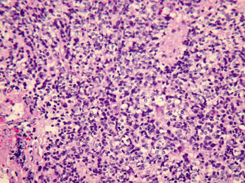
42+ Homer Wright Rosettes Seen In Medulloblastoma Background. Unfortunately, perivascular pseudorosettes are also less specific in that they are also encountered in medulloblastomas, pnets, central neurocytomas, and less often in glioblastomas, and a rare pediatric. A homer wright rosette is commonly seen in the following neurologic tumors:
Posterior Fossa Intra Axial Tumors Radiology Key from radiologykey.com
It consists of a halo of tumor cells surrounding a central region containing neuropil. A homer wright rosette is commonly seen in the following neurologic tumors: See what questions a doctor would ask.
They were mitotically active and.
Pseudorosettes because they contain fibrillary material. A homer wright rosette is commonly seen in the following neurologic tumors: Homer wright rosettes are considered pseudo in the sense that they are not true rosettes. Homer wright rosette — a circular or spherical grouping of dark tumor cells around a pale, eosinophilic, central area that contains neurofibrils but lacks a lumen;